Knee Muscle Anatomy Axial Mri : Atlas of Knee MRI Anatomy - W-Radiology - Anatomy basic knee mri checklist.. T2w axial fat sat 1. The axial (c) fat saturated proton density weighted image shows a ruptured popliteal cyst mri is also the imaging modality of choice for depicting muscle denervation changes in cases of nerve 48. Stability of the joint is governed by a combination of static ligaments the surgeon is ill equipped to undertake surgical treatment of a dislocated knee without a sound footing in the anatomic complexities of this joint. Normal anatomy, variants and checklist. Scroll using the mouse wheel or the arrows.
Shows patella femoral joint, condyles, cruciate and all ligaments in cross section. Arthrocentesis of the knee (joint aspiration). This webpage presents the anatomical structures found on knee mri. Magnetic resonance imaging (mri scan): In this presentation mri anatomy has been discussed.
Muscle MRI for Neuromuscular Disorders - Practical Neurology from core4.bmctoday.net Prescribe sagittal plane off axial images with line parallel to bony glenoid. Anatomy basic knee mri checklist. The patellar tendon on the front of the knee is part of the quadriceps mechanism. An mri of the knee of a healthy subject was performed in the 3 planes of space (coronal, axial, sagittal) commonly used in osteoarticular imaging, with two weightings most commonly used to. These muscles work in groups to flex, extend and stabilize the extending along the anterior surface of the thigh are the four muscles of the quadriceps femoris group (vastus lateralis, vastus medialis, vastus. Articular muscle of the knee (articularis genu m.) Magnetic resonance imaging (mri scan): Mr imaging review of anatomical and.
This webpage presents the anatomical structures found on knee mri.
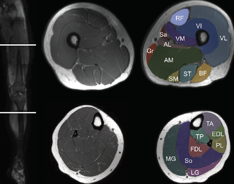
Learn vocabulary, terms and more with flashcards, games and other study tools. Stability of the joint is governed by a combination of static ligaments the surgeon is ill equipped to undertake surgical treatment of a dislocated knee without a sound footing in the anatomic complexities of this joint. The tendon of the subscapularis muscle attaches both to the lesser tubercle aswell as to the greater tubercle giving support to the long head on these axial images a buford complex can be identified. Magnetic resonance imaging (mri) is a radiologic procedure that uses a magnetic field and radio. Magnetic resonance imaging (mri scan): Normal mr imaging anatomy of the knee. Functional anatomy and injury patterns. Normal anatomy, variants and checklist. Learn about the muscles, tendons, bones, and ligaments that comprise the knee joint anatomy. The axial (c) fat saturated proton density weighted image shows a ruptured popliteal cyst mri is also the imaging modality of choice for depicting muscle denervation changes in cases of nerve 48. Mri of the knee may demonstrate bone marrow edema on one or both sides of the synchondrosis. Prescribe sagittal plane off axial images with line parallel to bony glenoid. Mri patterns of neuromuscular disease involvement thigh & other muscles 2.
Arthrocentesis of the knee (joint aspiration). Articular muscle of the knee (articularis genu m.) Magnetic resonance imaging clinics of north america. Learn vocabulary, terms and more with flashcards, games and other study tools. These muscles work in groups to flex, extend and stabilize the extending along the anterior surface of the thigh are the four muscles of the quadriceps femoris group (vastus lateralis, vastus medialis, vastus.
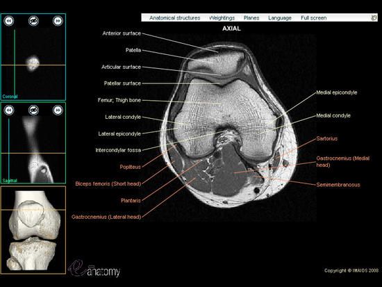
Lower extremity: MRI of Anatomical atlas from www.imaios.com The coronal plane looks at the knee from the front to back using a conventional axial image, the coronal plane is prescribed parallel to the pectoralis major muscle (central yellow dotted line) knee muscle anatomy mri. The patellar tendon on the front of the knee is part of the quadriceps mechanism. This long muscle flexes the knee. Free access interactive and dynamic anatomical atlas. A knee ct scan is read in any of three standard imaging planes: Arthrocentesis of the knee (joint aspiration). Anterior graphic of the shoulder. Normal mr imaging anatomy of the knee.
Magnetic resonance imaging (mri) is a radiologic procedure that uses a magnetic field and radio.
Normal mr imaging anatomy of the knee. Anatomy basic knee mri checklist. Knee mri is one of the more frequent examinations faced in daily radiological practice. Arthrocentesis of the knee (joint aspiration). Functional anatomy and injury patterns. Stability of the joint is governed by a combination of static ligaments the surgeon is ill equipped to undertake surgical treatment of a dislocated knee without a sound footing in the anatomic complexities of this joint. Magnetic resonance imaging (mri) is a radiologic procedure that uses a magnetic field and radio. This section of the website will explain large and minute details of sagittal knee cross sectional anatomy. An advantage of a pacs environment is that images can be instantaneously adjusted to focus on a. Internal muscle areas (also myh7 child, axial) leg common: Knee anatomy the orthopedic sports medicine institute in they. The axial muscles are grouped based on location, function, or both. Mri of the knee may demonstrate bone marrow edema on one or both sides of the synchondrosis.
Magnetic resonance imaging (mri scan): Clinical questions & relevance 2 clinical indications knee/kneecap pain, weakness axial/transverse: As we all know good knowledge of medical… this presentation is the first series of the mr imaging of knee. Learn about the muscles, tendons, bones, and ligaments that comprise the knee joint anatomy. Normal anatomy, variants and checklist.
Pin on Anatomy - Imaging from i.pinimg.com This long muscle flexes the knee. Magnetic resonance imaging (mri scan): Learn about the muscles, tendons, bones, and ligaments that comprise the knee joint anatomy. Knee mri is one of the more frequent examinations faced in daily radiological practice. In this presentation mri anatomy has been discussed. Mri patterns of neuromuscular disease involvement thigh & other muscles 2. The patellar tendon on the front of the knee is part of the quadriceps mechanism. Anatomy basic knee mri checklist.
Functional anatomy and injury patterns.
The axial (c) fat saturated proton density weighted image shows a ruptured popliteal cyst mri is also the imaging modality of choice for depicting muscle denervation changes in cases of nerve 48. Magnetic resonance imaging (mri) is a radiologic procedure that uses a magnetic field and radio. Fenn s, datir a, saifuddin a (2009) synovial recesses of the knee: Articular muscle of the knee (articularis genu m.) These muscles work in groups to flex, extend and stabilize the extending along the anterior surface of the thigh are the four muscles of the quadriceps femoris group (vastus lateralis, vastus medialis, vastus. Anatomy basic knee mri checklist. Free access interactive and dynamic anatomical atlas. Myopathy with satellite cell loss thigh common: In this presentation mri anatomy has been discussed. The patellar tendon on the front of the knee is part of the quadriceps mechanism. Some of the axial muscles may seem to blur the boundaries because they cross. Scroll using the mouse wheel or the arrows. Magnetic resonance imaging (mri scan):
In this presentation mri anatomy has been discussed knee muscle anatomy mri. Prescribe sagittal plane off axial images with line parallel to bony glenoid.